Anatomy of Flowering Plants
Habish Ribin Haneef
Updated on
Share:

Simplify your workflow in minutes.

Simplify your workflow in minutes.

Simplify your workflow in minutes.

Simplify your workflow in minutes.
Flowers, one of the most beautiful things in this world, right? People who do not love flowers’ beauty and fragrance may be rare. But have you ever imagined what these exquisite things constitute? The internal structure of flowering plants is a world of vast and enormous features. Anatomy is the study of the internal structure of organisms. The anatomy of flowering plants comprises organization and structure of tissues. So, to know the anatomy of flowering plants, first of all, you need to know what tissue is. In this blog, let’s briefly discuss the anatomy of flowering plants.
Table of Contents
ToggleThe meristematic tissue is made up of a group of similar and immature cells that have the capability to divide and form new cells. Meristematic tissue is again classified into three- apical meristem, intercalary meristem, and lateral meristem.
The Apical meristem is the primary meristem. The apical meristem occurs at the tips of roots and shoots. It increases the length of the plant.
The intercalary meristem is the meristem that develops between regions of mature or permanent tissues, especially in grasses. The intercalary meristem is capable of forming branches and flowers. Both the apical meristem and intercalary meristem are primary meristems.
The lateral meristem is the secondary meristem and is responsible for the secondary growth of the plant. The lateral meristem occurs in the mature regions of roots and shoots. The primary lateral meristem is the intrafascicular cambium and the secondary lateral meristems are the vascular cambium and cork cambium.
The permanent tissue is made up of structurally and functionally specialized cells which have lost the ability to divide. These tissues are derived from meristematic tissues. The permanent tissue is classified into two- simple permanent tissue and complex tissue.
Simple permanent tissues are those tissues having all the cells similar in structure and function. On the other hand, complex tissues are those tissues having different kinds of cells. Simple permanent tissues are again divided into three- parenchyma, collenchyma, and sclerenchyma.
Parenchyma is made up of thin-walled isodiametric cells. Their cell wall is made up of cellulose. In each cell, there is a large central vacuole and a peripheral cytoplasm containing a nucleus. Parenchyma is found in soft and non-woody areas of roots, stems, leaves, flowers, and fruits. The functions of parenchyma include photosynthesis, storage, and secretion.
Collenchyma is made up of living, closely packed isodiametric cells. Collenchyma consists of thickened cells at the corner. This thickening is due to cellulose, hemicellulose, and pectin. It is the collenchyma that provides mechanical support to the growing parts of the plant such as young stem.
Sclerenchyma is made up of dead cells with thick and lignified walls. Sclerenchyma are of two types- fibres and sclereids. Like collenchyma, sclerenchyma too provide mechanical support but it provides mechanical support to the mature plant organs.
Complex tissues are again divided into two- xylem and phloem.
Xylem is made up of tracheids, vessels, xylem fibre, and xylem parenchyma. Xylem conducts water and minerals from roots to other parts of the plant. Xylem is found deep in the plant and it provides mechanical strength.
Phloem is made up of sieve tube, companion cells, phloem parenchyma, and phloem fibres. Phloem transports food materials from leaves to various parts of the plant. Phloem is found towards the outer side.

The tissue systems are the structural and functional units of the plants. As you know tissue refers to a group of cells that share a common origin and perform the same function. Tissue systems refer to large units of tissues having some characteristics in common. The tissue system is classified into three- epidermal tissue system, ground tissue system, and vascular tissue system.
The epidermal tissue system is the outermost covering of the entire plant body and it comprises of epidermis, epidermal hairs, stomata, cuticle, root hairs, and trichomes. The epidermis is single-layered and it contains waxy thick layers of cuticle to prevent water loss. Epidermis also constitutes a number of hairs and these hairs are called root hairs.
A root hair is a unicellular extension of an epidermal cell.
Trichomes are found on stems and they may be multicellular, branched, or unbranched. Trichomes prevent water loss due to transpiration.
Stomata are found in the epidermis of leaves. Gas exchange and transpiration are regulated by the stomata.
The ground tissue system is made up of parenchyma, collenchyma, and sclerenchyma. The ground tissue system consists of simple permanent tissues. All the tissue systems between the epidermis and vascular bundle constitute the ground tissue system.
The vascular tissue system consists of vascular bundles made up of xylem and phloem.
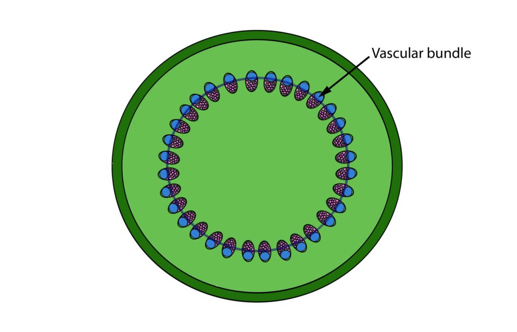
The vascular bundle is the part of the transport system in vascular plants comprised of xylem and phloem. Based on the arrangement of different parts, vascular bundles are classified into four- conjoint collateral vascular bundles, conjoint bicollateral vascular bundles, radial vascular bundles, and concentric vascular bundles.
In Conjoint collateral vascular bundles, the xylem and phloem are present on the same radius. It consists of a sequence of states. These types of vascular bundles appear in gymnosperms and angiosperms.
In Conjoint bicollateral vascular bundles, there are two phloem regions, one on each side of the xylem. It consists of two cambium belts, one on each side of the xylem.
In Radial vascular bundles, the xylem and phloem will be having different radii. In these vascular bundles, the pattern of development of the xylem is centripetal and hence radial vascular bundles are also called exarch.
Concentric vascular bundles are always closed and here either xylem is surrounding the phloem or the phloem is surrounding the xylem. Concentric vascular bundles are again divided into two- amphicribal or androcentric and ampivasal or lepto-centric. In Amphicribal vascular bundles, the phloem surrounds the xylem. The amphicribal vascular bundles are also called mesangial vascular bundles. They are found in ferns and lower gymnosperms. On the other hand, in Ampivasal vascular bundles, the xylem surrounds the phloem. They are also called endarch and are only found in angiosperms like Yucca and Dracaena.
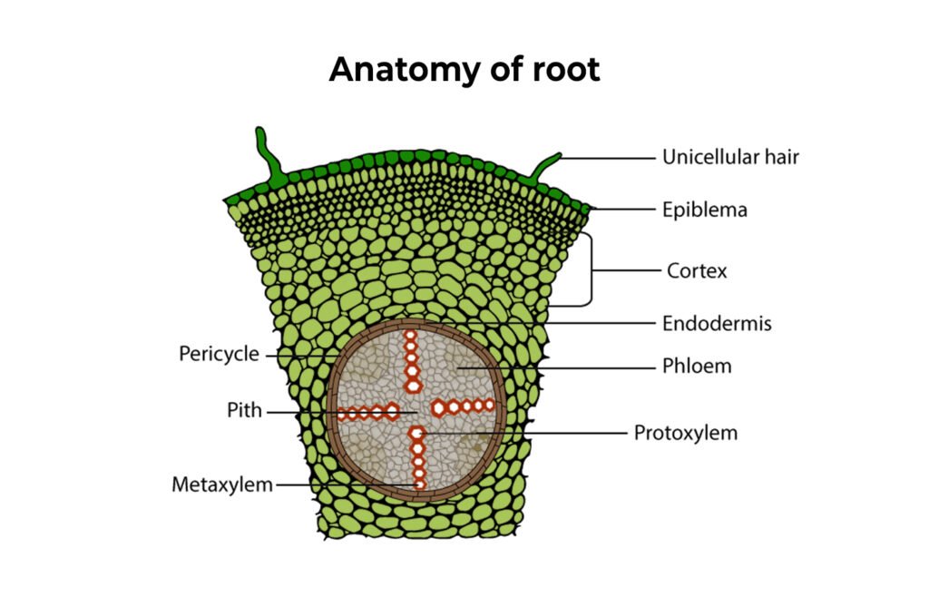
A root is an underground plant organ that is embedded in the ground. The root is developed from the radicle of the embryo. The primary functions of the root are to help in the anchorage of the plant and the conduction of water and minerals to all the parts of the plant. The anatomy of the root mainly includes dicot root and monocot root.
The dicot root consists of the epidermis, cortex, endodermis, pericycle, vascular bundles, and pith.
The epidermis is the outermost layer of a single row. It consists of unicellular root hair produced by certain cells called Epiblema. The epiblema is also called Rhizodermis or the Piliferous layer.
The cortex is made up of homogeneous parenchyma cells. It is found between the epidermis and endodermis layers. The cortex cells are circular or polygonal in shape with intercellular spaces. Water flows easily through the cortex because of the thin walls and sufficient space between the cells.
The endodermis is located between the vascular tissue and the cortex. The endodermis contains cells called Casparian strips which are barrel-shaped that possess special thickenings of suberin on radial and tangential walls. But Casparian strips are absent in those cells of the endodermis that are found in front of the protoxylem. These cells are called passage cells or transfusion cells and they help in the passage of water from the cortex to the pericycle.
The pericycle is single-layered and is composed of a type of parenchyma called prosenchyma. During secondary growth, it is the pericycle that forms the cork cambium. Lateral roots grow from the pericycle and hence they are endogenous.
The pith is made up of parenchymatous cells with intercellular spaces located at the centre. It is flexible.
Similar to the dicot root, the monocot root too consists of the epidermis, cortex, endodermis, pericycle, vascular bundles, and pith.
The epidermis consists of epiblema, the outermost layer composed of parenchymatous cells. Due to the elongation of epidermal cells, root hairs originate and it is the root hairs that help in the absorption of water and mineral salts. Here, the cuticle and stomata are absent.
The cortex is homogeneous and multi-layered. The cortex contains parenchyma cells which enclose the intercellular spaces for the exchange of gases. The parenchyma cells store food. In older roots, the outermost layer of the cortex produces protective exodermis.
The endodermis is the innermost layer of the cortex. It contains barrel-shaped cells with Casparian strips. Within the endodermis, passage cells are found and these are thin suberized cells.
The pericycle can be single-layered or multi layered. In the case of monocot roots, there is no cambium and so in pericycle there occurs only the formation of lateral roots.
The vascular bundles are arranged in a radial form and consist of alternate xylem and phloem bands. Due to the absence of cambium, the vascular bundles are closed type. Here, the xylem is exarch type. There are more than 6 xylem bundles in monocots and this condition is called the polyarch condition. The phloem consists of sieve tubes, companion cells, and phloem fibres. In monocots, phloem parenchyma is absent.
In monocots, the pith is large and well-developed.
Stems hold the plants upright so that they can receive the essential air and sunshine. It is a known fact that without a connection between the roots and the leaves, plants cannot survive in the air high above the ground. The two important functions of the stem are to transport food from leaves to other parts of the plant and to transport water and minerals from roots to leaves. Furthermore, the stems can produce cones, flowers, leaves, or secondary stalks.
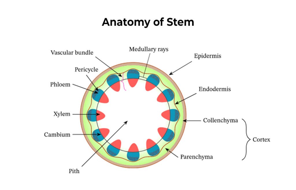
The dicot stem contains the epidermis, hypodermis, cortex, endodermis, pericycle, and vascular bundles.
The epidermis is the outermost layer of the stem. It is made up of a single layer of cells without chloroplast. The epidermis comprises of multicellular stem hair and stomata. It is the epidermis that provides protection.
The hypodermis is a thick layer made up of different kinds of cells. It is found under the epidermis. The hypodermis provides additional support to the epidermis. It consists of collenchyma which has chloroplasts and hence it is green and photosynthetic.
The cortex consists of parenchyma. The main function of the cortex is the storage of food.
The endodermis is the innermost membrane of the cortex. It is a single thick layer of cells. The endodermis cells are barrel-shaped. The function of these cells is to store starch in the stem of dicotyledonous trees and hence they are also called “starch sheath”.
The pericycle is located between the endodermis and vascular bundles, i.e., below the epidermis and above the vascular bundle. The pericyclic cells above the vascular bundle are made up of sclerenchyma and the remaining cells are made up of parenchyma. The pericycle present above the vascular bundle is called bundle cap.
The vascular bundles are composed of xylem and phloem. Each vascular bundle is an open conjoint collateral endarch. In the trunk of a dicotyledonous tree, the vascular bundles form a ring pattern. A well-developed pitch is present under the ring.
The anatomy of a monocot stem consists of the epidermis, hypodermis, ground tissue, vascular bundles, pith and stele.
The epidermis is the outermost layer and it is covered with thick skin bearing stomata but lacks multicellular hair.
The hypodermis is made up of 2-3 layers of sclerenchyma tissue.
The parenchyma cells near the hypodermis reaches the centre and are called ground tissue.
In ground tissue, the vascular bundles are present in scattered forms. Vascular bundles converging towards the center are larger and fewer in number. The peripheral vascular bundles are small but larger in number. The vascular tissue is surrounded by a layer of sclerenchyma tissue called bundle sheath.
Pith and stele– In monocots, a highly developed stele occurs called Atactostele.

Leaves are called “the kitchen of a plant” as photosynthesis occurs in leaves and so it is one of the crucial parts of a plant. Other than preparing food, leaves also significantly contributes to the carbon and oxygen cycle in the environment by producing oxygen during photosynthesis. The three primary parts of the leaves in flowering plants are the blade, petiole, and stipules. The three primary tissues of the leaves are the epidermis, mesophyll, and vascular tissue.
Secondary growth is the growth that results from cell division in the cambia or lateral meristems. This causes the stems and roots to thicken. Secondary growth occurs in most seed plants but it does not occur in monocots. It is a characteristic of dicotyledons and gymnosperms to form secondary vascular tissues from their cambia.
During secondary growth in dicot stem, an increase in diameter in plants occurs. Five important things also occur in the secondary growth of a dicot stem- formation of the cambial ring, formation of secondary xylem and secondary phloem from the cambial ring, formation of heart wood and sap wood, formation of spring wood and autumn wood, and development of cork cambium.
The secondary growth in dicot root takes place due to the activity of the vascular cambium. The vascular cambium is produced in the stele and cortex and ultimately results in the increase in the diameter of dicot roots.
Read more: How does an AC generator function?
The term tissue refers to a group of cells that share a common origin and usually perform the same function. Tissues are classified into two- Meristematic Tissue and Permanent Tissue.
Collenchyma provides mechanical support to the growing parts of the plant such as young stem.
All the tissue systems between the epidermis and vascular bundle constitute the ground tissue system.
Secondary growth is the growth that results from cell division in the cambia or lateral meristems.
The three primary parts of the leaves in flowering plants are the blade, petiole, and stipules.
The function of endodermis in a dicot stem is to store starch in the stem of dicotyledonous trees and hence they are also called “starch sheath”.