Eyes are termed as photoreceptors as they are chief organs of vision in the human body!
Bony sockets of the skull inside which human eyes are located are called orbits. The human eye which is in the form of a spherical ball is guarded by two eyelids: the upper eyelid and lower eye lid which are capable of moving frequently.
Hairs bordering the eyelids are called eye-lashes. The third eyelid which is vestigial is called plica semilunaris. It lies at the corner of the eye. Meibomian glands present on the margins of eyelids secrete an oily substance for lubricating the eyelids and for stocking a thin film of tears underneath.
Lacrimal or tear glands produce tears which keep the eyeball moist. They are present below the outer corner of the upper eyelid. Tears flow across the front of the eye. Excessive tears during emotional moments are drained into a small lacrimal sac at the inner margin. Afterwards they are discharged by means of nasolacrimal duct into the nasal passageways.
Tears are essential for washing away dust particles fallen on the eyeball.
They help in killing germs, thus preventing infection.
Tears help us communicate emotions.
Six extrinsic muscles help in moving the eyeball in the orbit. While the superior oblique muscle helps in downward movement , the inferior oblique muscle facilitates upward and outward movement. The medial rectus muscle is responsible for inward movement and internal rectus muscle for the outward movement. Similarly the superior rectus muscle is used for upward movement and inferior rectus in downward movement.
The eyeball consists of three coats. The outer sclerotic, middle choroid and the inner retina. While one end of the muscles are attached in the bony socket, the other end of the muscles are attached to the outer coat of the eyeball. They are responsible for the movement of the eyeball from side to side and up and down, helping us control the direction of vision.
Layers of Eye
Sclerotic: Formed of a tough layer of modified fibrous connective tissue, it is the outermost layer of the eyeball. It is transparent and non-vascular on the front side, known as cornea. Another transparent but vascular membrane called conjunctiva is present over the cornea. It is an extension of the skin of the eyelid. The sclerotic layer protects the eye, gives shape to the eyeball and also provides a surface for attachment of six extrinsic muscles.
Choroid: It is the middle layer. Choroid consists of highly vascular connective tissue with dark brown pigment. This layer in nocturnal mammals contains a silvery connective tissue (tapetum) for reflecting light. This causes their eyes to shine at night.It thickens as a circular ciliary body in the front.
Choroid is made up of blood vessels, glands and ciliary muscles. Choroid separates from the sclerotic in front of the ciliary body and passes inwards as Iris. The iris possesses a circular aperture in the center called pupil. Behind the Iris, we have a transparent biconvex lens, it is attached to the ciliary body by suspensory ligaments.
The cavity of the eyeball is divided into a small, anterior aqueous chamber and a large, posterior vitreous chamber by the iris and lens. The vitreous chamber is filled with gelatinous vitreous humor while the aqueous chamber is filled with watery aqueous humor. The aqueous humor consists of 98 percent water, protein and sodium chloride.
Choroid maintains intraocular pressure. It acts as a refractive medium which supplies nutrition to the lens and helps in draining away metabolic wastes. Vitreous and aqueous humors have similar composition. But vitreous humor contains less glucose and higher concentration of pyruvic acid and lactic acid. Lymphatic vessels are present in the vitreous chamber. It passes from the lens to the blind, and is called the hyaloid canal.
Retina: Retina is the innermost layer of the eyeball. It consists of two sub-layers.
(a) Outer layer of pigment cells. They can be found lying immediately after the choroid.
(b) Rods and cones
The rods can distinguish between various degrees of light and darkness, it contains rhodopsin pigment or visual purple. Nocturnal animals are able to see in the dim light as their retina are mainly formed of this pigment. Cones consist of iodopsin pigment and cyanopsin which deals with color vision.
The three other recently discovered pigments in the human eye are erythrolabe, chlorolabe, and cyanolabe. They are sensitive to red, green, and blue light respectively. The pigments are located in cones and are responsible for color vision.
Rods are cylindrical while cones are pyramidal in shape. They are not evenly distributed in the retina. The shape of the cones vary in different parts of the retina. An eye contains 100 million rods and about 7 million cones. Yellow spot or area centralis contains a large number of cones.
The area centralis is a small depression, it is the area of the sharpest color vision. The blind spot lies behind the yellow spot. It lacks both rods and cones. The blind spot lacks rods and cones. The optic nerve arises from the blind spot.
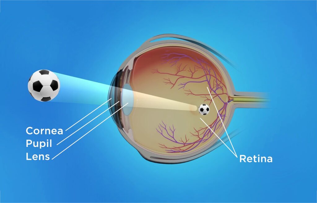
Working of the eye
Before falling on the biconvex lens, the light rays from an object pass through the conjunctiva, cornea and the pupil. The amount of light that will enter into the eyeball is determined by the size of the pupil. When the intensity of light is low, the size of the pupil enlarges. On the other hand, the size diminishes when the intensity is high.
The biconvex lens focuses light rays on the retina, forming a sharp inverted image on the retina. The optic nerve carries the impression of the inverted retinal image to the brain, where it gets interpreted. By adjusting the focal length of its lens, the eye helps us in seeing objects far away and nearby. This ability is called the power of accommodation of the eye.
In animals, the lens is normally focussed for viewing distant objects. The curvature of the lens is reduced while viewing nearby objects.
The fields of vision of two eyes overlap or even coincide in higher mammals including human beings. This is called binocular vision. But in the case of animals like rabbits, each eye covers a different field of vision. We call this monocular vision.
Some common eye defects
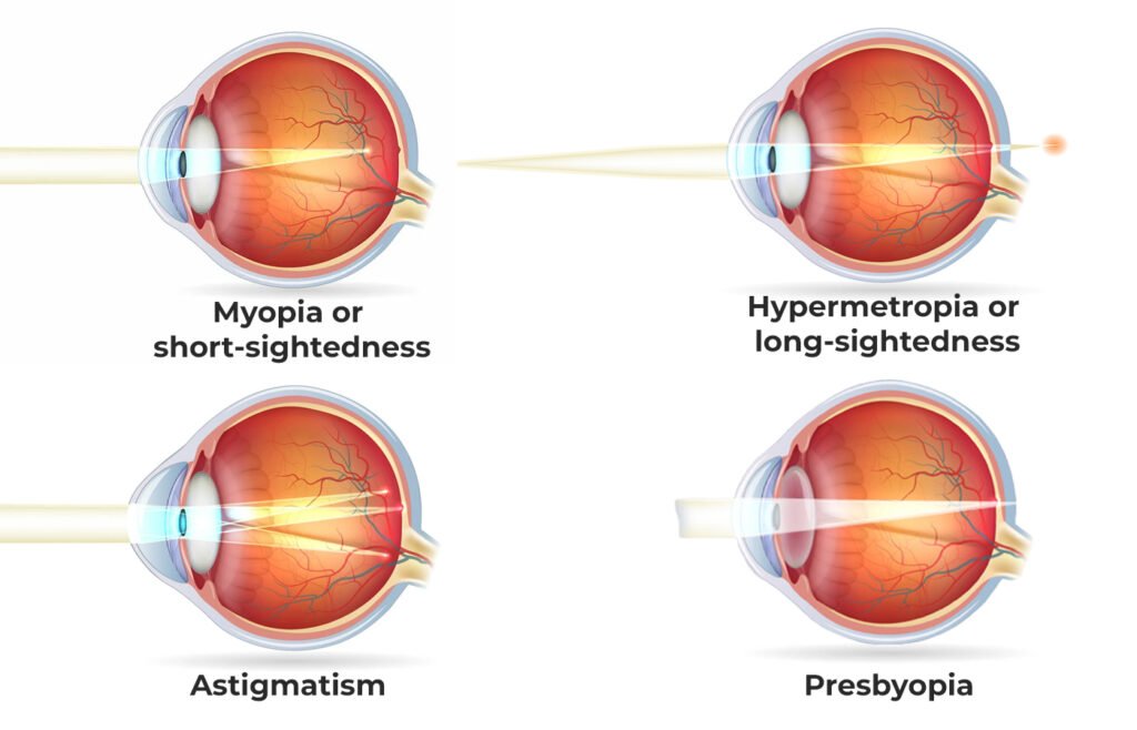
Myopia or short-sightedness
People who have this eye defect can see nearby objects clearly, but not objects at distance. In this condition, the lens of the eye is too convex. The rays of light get focussed at a point in front of the retina, and not upon it. The defect is corrected using a concave lens.
Hypermetropia or long-sightedness
In this case a person will be able to see distant objects clearly but not objects nearby. The eye ball will be too short which makes the retina too close to the lens. The focussing point will lie behind the retina. The condition is corrected using a convex lens.
Astigmatism
Irregularities in the shape of lens and cornea cause astigmatism. In different regions of the eye, the lens will have different curvatures. Due to this the light rays cannot be brought into the sharp focus of the retina. The defect can be corrected using a cylindrical lens.
Presbyopia
Loss of flexibility of the lens in old age causes presbyopia. The person will find it difficult to focus on near objects. The defect can occur anytime after the age of 35. Old-age Sight can be corrected using convex lenses.
Cataract
Cataract in an eye defect in which the lens becomes opaque due to several factors. In this case a person won’t be able to see objects clearly as light rays won’t pass through the lens properly. The only remedy for cataract is surgical removal of the lens and its replacement by a convex lens.
Glaucoma
The pressure within the eye rises above normal value – 15-20 mm. In this abnormality, the intraocular tension ultimately causes blindness.