Heart is the organ which pumps blood throughout the body through the vessels of the circulatory system. This helps in supplying oxygen and nutrients to the tissues and also helps in removing carbon dioxide and other wastes.
The heart is located in the center of our chest. It is placed slightly to the left of the sternum/breastbone. The heart sits between our lungs and is encased in a double walled sac known as the pericardium.The pericardium helps in protecting the heart and anchoring it inside the chest. Pericardial fluid acts as a lubricant. It lubricates the heart during contractions and movements of the lungs and diaphragm.
The human heart is approximately the size of a large fist. It weighs somewhere between 280-340 grams in men and 230-280 grams in women.
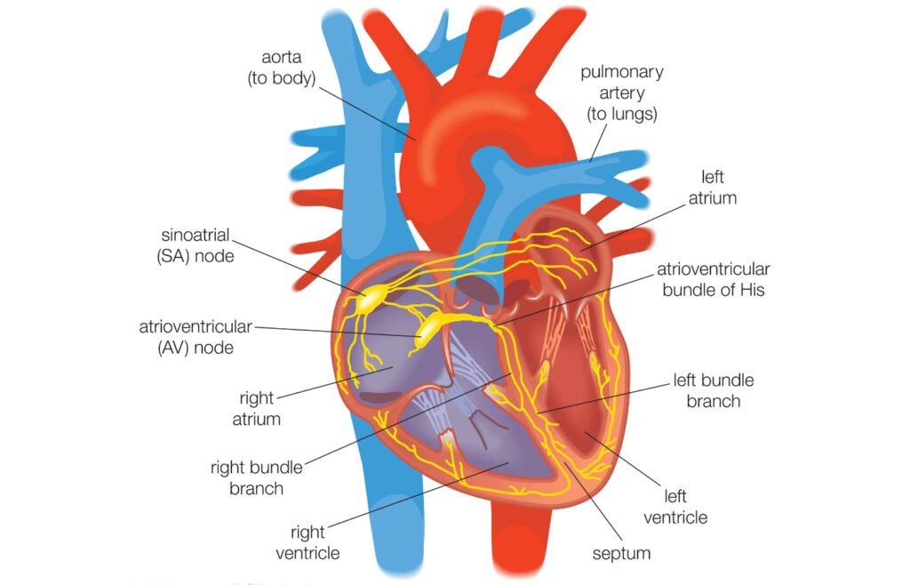
The human heart consists of four chambers. The atria – the two upper chambers and the ventricles – the two lower ones. The right atrium and the right ventricle comprise the “right heart”, whereas the left atrium and the left ventricle constitutes the “left heart”. The wall of muscle which separates two sides of the heart is known as the septum.
The heart’s outer wall has three layers. The outermost wall layer is known as the epicardium, the middle layer is called the myocardium, it contains the muscle which contracts the heart. The heart chambers are lined by inner layers/endocardium.
The upper and lower chambers of the heart are connected by the atrioventricular (AV) valves. The right ventricle is separated from the pulmonary artery by the pulmonary semi-lunar valve. On the other hand, the left ventricle is separated from the aorta by the aortic valve. The valves are anchored to heart muscles by heartstrings/chordae tendinae.
How does the human heart function?
Blood is circulated by the heart through two pathways, they are known as the pulmonary circuit and the systemic circuit.
Pulmonary Circuit
The deoxygenated blood leaves the right ventricle of the heart through the pulmonary artery and moves to the lungs. Following this, the oxygenated blood returns through the pulmonary vein and enters the left atrium of the heart.
Systemic Circuit
Oxygenated blood leaves the heart and moves through the left ventricle and reaches the aorta. From there blood enters the arteries and capillaries for supplying oxygen to body tissues. Deoxygenated blood which returns through veins finally re-enters the heart’s right atrium.
While the left main coronary artery branches into the left anterior descending artery and the left circumflex artery, the right coronary artery is spread out on the right side of the aorta.
Blockage of any of these arteries can lead to a heart attack, or can cause damage to the heart muscle. A heart attack is distinct from what we call a cardiac arrest. The latter can be defined as a sudden loss of heart function which usually occurs due to electrical disturbances of the heart rhythm. A heart attack may lead to cardiac arrest, but a cardiac arrest can happen due to several other reasons as well.
Our heart contains electrical “pacemaker” cells which helps in producing heartbeats.
Valves are responsible for preventing backflow, it helps to keep the blood flow through one direction through the heart.
Interestingly, every single day, our heart beats around 100,000 times, pumping approximately 6.8 liters of blood per minute through the 60,000 miles of blood vessels which are present in our body!
In this blog we have discussed the anatomy and functions of the human heart.